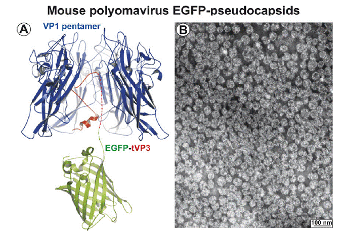
Structure and architecture of the analyzed EGFP capsid-like particles derived from mouse polyomavirus. (A) View-through, showing the macromolecular interaction of VP1 pentamer (depicted in blue) with the C terminus of minor structural protein VP3 (depicted in red) fused with the EGFP protein (depicted in green). (B) Electron microscopy: EGFP-VLPs purified frominsect cell lysates by density gradient were attached to carbon-coated grids, contrasted by negative staining and visualized by electron microscopy. Detailed design, production, properties, and characterization of chimeric EGFP-VLPs were described by Boura et al. 2005.