Exobiology Group
Institute of Geochemistry, Mineralogy and Mineral Resources
Faculty of Science, Charles University
News
30. 8. 2024
Microbial colonization of gypsum: from the fossil record to the present day

Microorganisms inhabiting gypsum have been observed in environments that differ greatly in water availability. Gypsum colonized by microorganisms, including cyanobacteria, eukaryotic algae, and diverse heterotrophic communities, occurs in hot, arid or even hyperarid environments, in cold environments of the Antarctic and Arctic zones, and in saline and hypersaline lakes and ponds where gypsum precipitates. Fossilized microbial remnants preserved in gypsum were also reported. Gypsum protects the endolithic microbial communities against excessive insolation and ultraviolet radiation, while allowing photosynthetically active radiation to penetrate through the mineral substrate. We here review the worldwide occurrences of microbially colonized gypsum and the specific properties of gypsum related to its function as a substrate and habitat for microbial life on Earth and possibly beyond. Methods for detecting and characterizing endolithic communities and their biomarkers in gypsum are discussed, including microscopic, spectroscopic, chemical, and molecular biological techniques. The modes of adaptation of different microorganisms to life within gypsum crystals under different environmental conditions are described. Finally, we discuss gypsum deposits as possible targets for the search for microbial life or its remnants beyond Earth, especially on Mars, where sulfate-rich deposits occur, and propose strategies to detect them during space exploration missions. → Link to article at journal webpage.
30. 7. 2024
Comparative analysis of cyanobacterial communities in gypsum outcrops: insights from sites in Israel and Poland

Today, the biodiversity of endolithic microbial colonisations are only partly understood. In this study, we used a combination of molecular community metabarcoding using the 16S rRNA gene, light microscopy, CT-scan analysis, and Raman spectroscopy to describe gypsum endolithic communities in 2 sites—southern Poland and northern Israel. The obtained results have shown that despite different geographical areas, climatic conditions, and also physical features of colonized gypsum outcrops, both of these sites have remarkably similar microbial and pigment compositions. Cyanobacteria dominate both of the gypsum habitats, followed by Chloroflexi and Pseudomonadota. Among cyanobacteria, Thermosynechococcaceae were more abundant in Israel while Chroococcidiopsidaceae in Poland. Interestingly, no Gloeobacteraceae sequences have been found in Poland, only in Israel. Some of the obtained 16S rRNA gene sequences of cyanobacteria matched previously detected sequences from endolithic communities in various substrates and geographical regions, supporting the hypothesis of global metacommunity, but more data are still needed. Using Raman spectroscopy, cyanobacterial UV-screening pigments—scytonemin and gloeocapsin have been detected alongside carotenoids, chlorophyll a and melanin. These pigments can serve as potential biomarkers for basic taxonomic identification of cyanobacteria. Overall, this study provides more insight into the diversity of cyanobacterial endolithic colonisations in gypsum across different areas. → Link to article at journal webpage.
14. 6. 2024
Evaluation of pore-fracture microstructure of gypsum rock fragments using micro-CT

This study utilized X-ray micro-computed tomography (micro-CT) to investigate weathered gypsum rocks which can or do serve as a rock substrate for endolithic organisms, focusing on their internal pore-fracture microstructure, estimating porosity, and quantitative comparison between various samples. Examining sections and reconstructed 3D models provides a more detailed insight into the overall structural conditions within rock fragments and the interconnectivity in pore networks, surpassing the limitations of analyzing individual 2D images. Results revealed diverse gypsum forms, cavities, fractures, and secondary features influenced by weathering. Using deep learning segmentation based on the U-Net models within the Dragonfly software enabled to identify and visualize the porous systems and determinate void space which was used to calculate porosity. This approach allowed to describe what type of microstructures and cavities is responsible for the porous spaces in different gypsum samples. A set of quantitative analysis of the detected void and modeled networks provided a needed information about the development of the pore system, connectivity, and pore size distribution. Comparison with mercury intrusion porosimetry showed that both methods consider different populations of pores. In our case, micro-CT typically detects larger pores (> 10 μm) which is related to the effective resolution of the scanned images. Still, micro-CT demonstrated to be an efficient tool in examining the internal microstructures of weathered gypsum rocks, with promising implications particularly in geobiology and microbiology for the characterization of lithic habitats. → Link to article at journal webpage.
10. 3. 2023
Using Raman spectroscopy to detect scytonemin of epiliths and endoliths from marble, serpentinite and gypsum
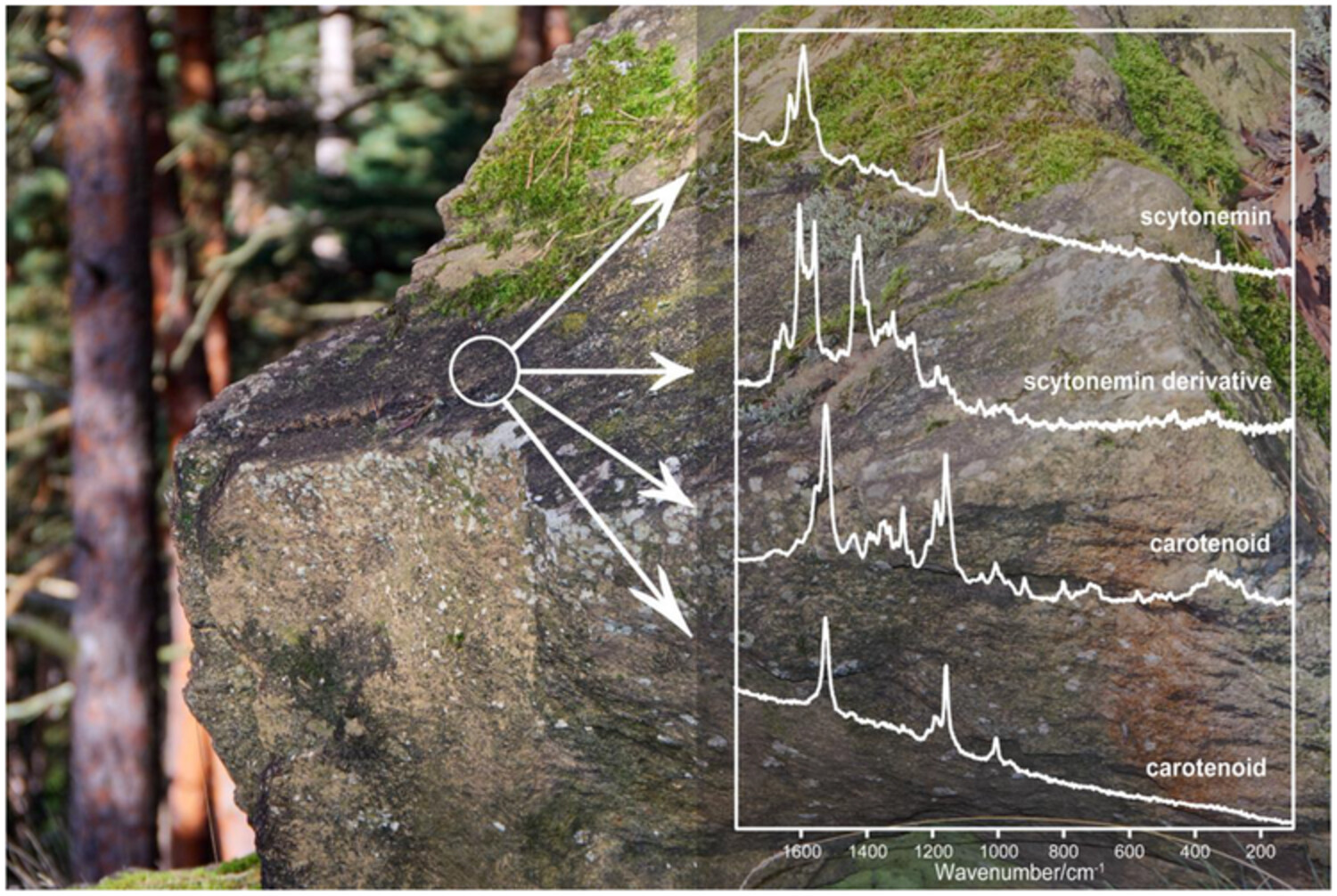
Here, we present Raman spectra showing the presence and distribution of scytonemin and carotenoids in epilithic and endolithic colonisations from temperate locations in Central Europe and Sicily. In the Bohemian Massif, marble and serpentinitic cyanobacterial epiliths dominated by cyanobacteria Scytonema, Stigonema, Hassallia, Gloeocapsopsis and Gloeocapsa were investigated using light microscopy and Raman spectroscopy. Scytonemin was a common dark pigment, accompanied by carotenoids and gloeocapsin on the marbles from Opolenec and on serpentinites from Holubov (South Bohemia). Raman spectra from other sites originated from endolithic colonisations of gypsum. They were located in the Carpathian foredeep (Badenian, Silesian unit, eastern Poland) and in Messinian complexes in the Mediterranean area (Sicily). Similarly to the previous localities, almost ubiquitous occurrence of scytonemin confirmed the presence of cyanobacterial colonisations. Obtained findings are important from the spectroscopic point of view. Additionally, comparing results from several sites confirmed the common occurrence of scytonemin in both endoliths and epiliths from areas that cannot be considered climatically extreme, although they experience rapid fluctuations in temperature, humidity and UV irradiation on the exposed rocky substrates. → Link to article at journal webpage.
14. 2. 2023
Scytonin in gypsum endolithic colonisation: First Raman spectroscopic detection of a new spectral biosignature for terrestrial astrobiological analogues and for exobiological mission database extension
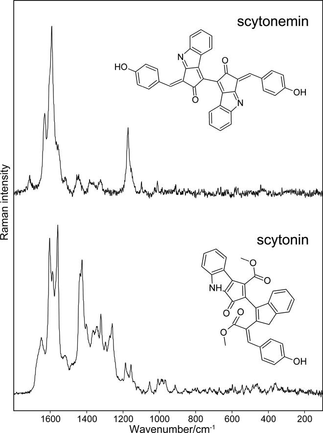
Microbial colonisations of gypsum from Eastern Poland (Badenian, Middle Miocene age) were investigated by Raman microspectrometry with a rarely used excitation 445 nm excitation. Zones of microbial colonisation in selenitic gypsum endolithic outcrops comprise algae and cyanobacteria, which commonly contain the photosynthetic and protective pigments carotenoids, scytonemin and gloeocapsin. Diagnostic bands differing from those of scytonemin have been identified in black colonies in gypsum outcrops at Chotel Czierwony (Poland). Raman spectral signatures of scytonin are reported here for the first time in two endolithic specimens identified by the band wavenumbers predicted from DFT calculations. The strong or medium strong intensity Raman bands observed at 1603, 1585, 1559, 1435, and 1424 cm−1. Other weaker bands were located at 1676 (sh), 1660 (sh), 1649, 1399, 1362, 1342, 1320, 1294, 1272, 1259, and 1052 cm−1. The first observation of the Raman spectrum of scytonin in the cyanobacterial colonisation of gypsum facilitates the inclusion of this new biomolecular signature in the library of unique Raman spectra of biological pigments invaluable for detection of traces of life in frame of the planetary missions. → Link to article at journal webpage.
23. 8. 2022
Critical evaluation of portable Raman spectrometers: From rock outcrops and planetary analogs to cultural heritage – A review
Review article

This review focuses on the progress made and recent developments regarding the use of portable Raman spectrometric devices in the geosciences as well as with scientific testing of objects of cultural heritage. By utilizing different types of Raman spectrometric devices - from the modular; through systems operating with optical heads mounted via optical fibers; to the compact, lightweight, and handheld - are herein described and critically evaluated. The wide range of applications under indoor and closed environments (museums, collections, religious buildings, etc.) are reviewed, and the more commonly used for characterization of art in the broadest sense, as well as fully outdoor investigations (e.g., on rocky outcrops; of the minerals, microbiological colonization, etc.), and including extreme Earth environments and projects with exo-planetary research potential. The rapid acquisition of relevant spectroscopic data and the non-destructiveness of the analyses and diagnostics are greatly appreciated by art historians and conservators on the one hand, as well as by mineralogists, geoscientists, microbiologists on the other. The various environments investigated and the broad range of compounds that can be detected by obtaining their Raman spectra have demonstrated the vast potential of portable, and especially handheld, Raman spectrometer devices for on-site applications. → Link to article at journal webpage.
24. 3. 2022
The GeoRaman 2022 Conference will be held in Prague: 29 August to 2 September
![]()
15TH INTERNATIONAL GEORAMAN CONFERENCE GeoRaman 2022 Prague. We are delighted to announce that, after a forced interruption due to the pandemic, the 15th edition of the international GeoRaman conference will return in 2022. The conference will be held in the marvellous city of Prague, Czech Republic, for the second time in 20 years, from the 29th of August to the 2nd of September 2022. The GeoRaman conference will cover topics from research areas of Raman spectroscopy applications in geology and mineralogy, cultural heritage, and exobiology. A field trip will be a part of the conference. 15th International GeoRaman Conference: GeoRaman 2022 Prague 29 August to 2 September
11. 4. 2022
Analysis of brown, violet and blue pigments of microorganisms by Raman spectroscopy
Review article
![]()
Raman spectroscopy allows the detection and discrimination between microbial pigments of different origins, including carotenoids, chlorophylls and less commonly found violet and brown non-photosynthetic pigments. This review reports examples of Raman spectroscopic studies on violet and brown pigments produced by microorganisms. Examples of investigations on the biological pigments scytonemin, gloeocapsin, violacein, phycocyanin, and prodigiosin are reviewed. Raman spectroscopy is a fast, simple and direct analytical tool which has been appreciated by microbiologists and chemists but more recently also by those who study natural geobiological processes in real-world systems. Different Raman spectroscopic approaches including confocal Raman spectrometry, FT-Raman spectroscopic possibilities, and SERS modes are reviewed for possibilities of following distribution of pigments at the cellular level in microbial cultures or in native endoliths, including confocal Raman spectrometry, FT-Raman spectroscopic possibilities, and SERS modes. New fast Raman mapping techniques are increas-ingly used, as well as portable systems. Special possibilities of high relevance for geoscience, geobiology or applied microbiology are opened up by the recent revolutionary technical developments, including the availability of portable and handheld tools that can be used directly on rocky outcrops to study native microbial colonization in situ. → Link to article at journal webpage.
3. 9. 2021
Evaluation of miniaturized Raman spectrometers for planetary exploration: From aromatics to amino acids
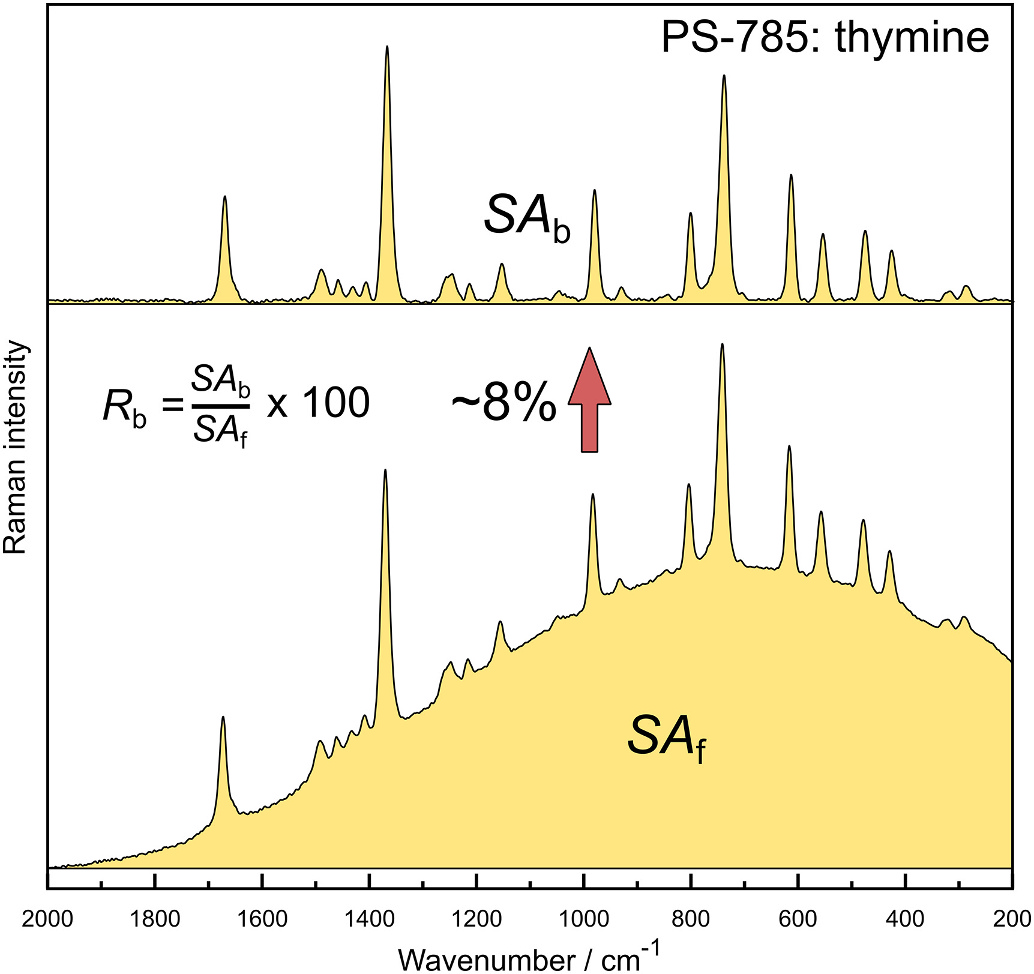
Organic molecules are currently believed to be abundant in space, but the possible biogenic origin, or the mere existence, on some planetary surfaces, Mars specifically, is a pending question. Reliable methods of detection are required to answer this question unambiguously and Raman spectroscopy has already been suggested for this task years ago. With exploration missions aiming to Mars on the horizon, collecting experience and building databases will have crucial importance investigations of analytical data obtained through Raman instrumentation onboard of rovers in the frame of Mars 2020 and other forthcoming missions. This work focuses on the evaluation of some portable Raman systems coupled to different excitation lasers (532, 785, 1064 nm and a dual laser system with sequentially shifted excitation SSE) for the detection of various organic molecules, with emphasis on non-complicated measure protocol and observation of fluorescence emission when a different wavelength is used. By using a simple statistical approach, we demonstrate a generally good readability of the obtained spectra for most of the investigated organics regardless the excitation sources and instruments used. A varying level of fluorescence emission was encountered, resulting in higher background for the 532 nm and 785 nm instrumentation while 1064 nm and SSE spectrometers provided almost fluorescence-free spectra. These results illustrate how the relatively simple miniaturized Raman spectrometers can provide fast and unambiguous identification of various organic compounds which are of great importance in the current and future planetology and/or exobiology missions. → Link to article at journal webpage.
10. 7. 2021
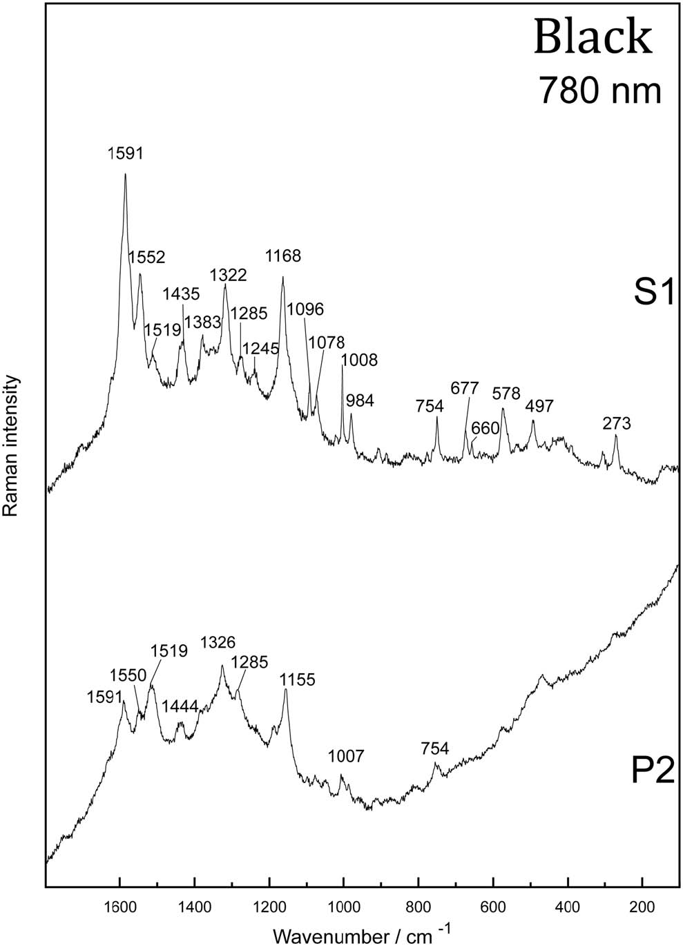
Raman spectroscopic search for scytonemin and gloeocapsin in endolithic colonizations in large gypsum crystals
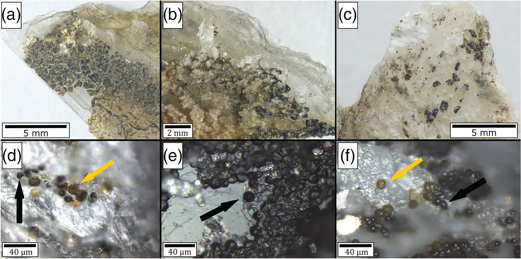
Rock-dwelling microorganisms are frequently exposed to intense UV-radiation, and thus, they synthesize specialized UV-protective pigments. In this study, Raman microspectrometry was used for analysis of noncarotenoid UV-protective pigments of dark-pigmented endolithic colonization found in gypsum outcrops from three areas (Sicily, Poland and Israel). Samples were investigated using 445-, 532- and 780-nm excitation lasers. Two noncarotenoid UV-protective pigments scytonemin and gloeocapsin were detected in all studied sites. Major Raman bands of scytonemin were found at around 1593, 1552, 1438 and 1173 cm-1. Gloeocapsin shows characteristic Raman bands similar to anthraquinone-based parietin of lichens: at 1665, 1575, 1378, 1310 and 465 cm-1. Scytonemin and gloeocapsin are highly specific for cyanobacteria and therefore can be used as biomarkers of certain taxa of cyanobacteria. Detection of such pigments by Raman spectroscopy using three excitation wavelengths allows to gather more information about the composition of endolithic consortia using relatively fast and noninvasive methods and in their natural habitats. → Link to article at journal webpage.
15. 6. 2021
Detecting pigments from gypsum endoliths using Raman spectroscopy: From field prospection to laboratory studies
Microbial colonisations of gypsum from different sites from Southern Sicily and Eastern Poland were investigated using laboratory-based Raman microspectrometers and portable Raman spectrometric devices. Selected zones of microbial colonisations of few types of gypsum are described from the point of view of the presence of algae and cyanobacteria. Macrocrystalline gypsum layers in Sicily and Eastern Poland originate from Tertiary sedimentary series. In Southern Sicily gypsum outcrops from late Miocene age were investigated near Scala dei Turchi, Torre Salsa and Siculiana Marina. Polish Tertiary Middle Miocene age examples of gypsum colonisations of decimetre to metre long outcropping gypsum crystals were studied near Chotel Czerwony, Skorocice and Chwalowice. Common microbial pigments were detected: carotenoids were documented in majority of the samples (common Raman bands at around 1525, 1157 and 1004 cm-1), Raman spectra of other pigments were recorded in several zones using near infrared excitation (785 nm): chlorophyll (1326, 1285, 1188 and 745 cm-1), scytonemin (1593, 1552, 1438 and 1173 cm-1) and phycobiliproteins (1275 cm-1). Raman microspectrometric investigations of colonisations allow to gather detailed information about pigment distribution in micrometric zones of gypsum samples. Portable instrumentation permits also detection of carotenoids. Observed shifts of positions of Raman features of carotenoids between gypsum samples (and sites worldwide) and relative to reference values are discussed and critically evaluated. → Link to article at journal webpage.
18. 2. 2021
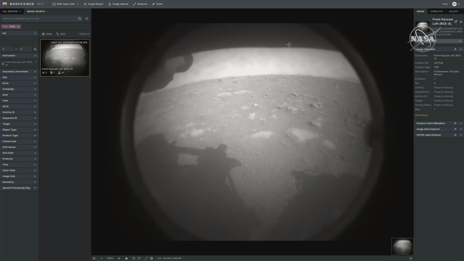
Perseverance has landed on Mars!
27. 4. 2020
Fast screening of carotenoids of gypsum endoliths using portable Raman spectrometer (Messinian gypsum, Sicily)
We tested the potential of a portable Raman spectrometer-RaPort (G) by EnSpectr with a 532-nm laser for use in field detection of biomarkers of endolithic colonization in gypsum. This study extends our previous research on gypsum endolithic colonies using laboratory Raman microspectrometer (514.5- and 785-nm lasers). In the current study, variously coloured layers were found and screened separately on the outcrop under natural conditions. In the recorded Raman spectra, stretching vibrations of carotenoids were observed - v1 (C=C) around 1,513 cm-1, and v2 (C-C) around 1,154 cm-1, as well as the feature at around 1,005 cm-1. The exact identification of different carotenoids by portable Raman spectrometer is limited. Nevertheless, the portable Raman spectrometer allowed fast and in situ detection of carotenoids of variously pigmented gypsum endolithic zones. Application of miniature Raman instruments is important in the context of forthcoming astrobiological missions to Mars (Exomars and Mars 2020). → Link to article at journal webpage.
1. 1. 2020
Raman spectroscopic screening of cyanobacterial chasmoliths from crystalline gypsum - The Messinian crisis sediments from Southern Sicily
This study shows the first results on Raman spectroscopic investigations of pigments from cyanobacterial endolithic colonisations in gypsum of Messinian age (Miocene). Gypsum from outcrops of sedimentary series close to Eraclea Minoa (Sothern Sicily) is sometimes inhabited by endoliths. Raman spectroscopic investigations allow to specify type of colonisation through identification of their pigments. Coccoid cyanobacteria (Chroococcidiopsis sp., Gloeocapsopsis pleurocapsoides, Gloeocapsa compacta, and Synechococcus sciophilus), filamentous heterocytous Nostoc sp., and filamentous bundle-forming Symplocastrum cf. aurantiacum and Microcoleus sp. were found to inhabit different parts of the gypsum samples. Carotenoids (as assumed frequently beta-carotene) are produced in all the samples studied (optimised detection using 514-nm excitation), scytonemin in the samples colonised by G. pleurocapsoides (G1, G2, and G6), and phycobiliproteins in the sample G3 (those detected using 785-nm excitation). This study is the first initiative to collect better knowledge on detecting biomarker traces in the frame of Messinian-age gypsum sequence of relevance for old Martian environments. → Link to article at journal webpage.
5. 12. 2019
Detection of carotenoids of halophilic prokaryotes in solid inclusions inside laboratory-grown chloride and sulfate crystals using a portable Raman spectrometer: applications for Mars exploration
Inclusions in evaporitic minerals sometimes contain remnants of microorganisms or biomarkers, which can be considered as traces of life. Raman spectroscopy with resonance enhancement is one of the best analytical methods to search for such biomarkers in places of interest for astrobiology, including the surface and near subsurface of planet Mars. Portable Raman spectrometers are used as training tools for detection of biomarkers. Investigations of the limits and challenges of detecting biomolecules in crystals using Raman spectroscopy is important because natural occurrences often involve mineral assemblages as well as their fluid and solid inclusions. A portable Raman spectrometer with 532 nm excitation was used for detection of carotenoid biomarkers: salinixanthin of Salinibacter ruber (Bacteroidetes) and a-bacterioruberin of Halorubrum sodomense (Halobacteria) in laboratory-grown artificial inclusions in compound crystals of several chlorides and sulfates, simulating entrapment of microorganisms in evaporitic minerals. Crystals of halite (NaCl), sylvite (KCl), arcanite (K2SO4) and tschermigite ((NH4)Al(SO4)2 · 12H2O) were grown from synthetic solutions that contained microorganisms. A second crystalline layer of NaCl or K2SO4 was grown subsequently so that primary crystals containing microorganisms are considered as solid inclusions. A portable Raman spectrometer with resonance enabling excitation detected signals of both carotenoid pigments. Correct positions of diagnostic Raman bands corresponding to the specific carotenoids were recorded. → Link to article at journal webpage.
29. 5. 2019
Comparison of Miniaturized Raman Spectrometers for Discrimination of Carotenoids of Halophilic Microorganisms

We present a comparison of the performance of four miniature portable Raman spectrometers for the discrimination of carotenoids in samples of carotene-producing microorganisms. Two spectrometers using a green laser allowing to obtain Resonance Raman (or pre-Resonance Raman) signals, one instrument with a 785 nm laser, and a recently developed Portable Sequentially Shifted Excitation Raman spectrometer (PSSERS) were used for identifying major pigments of different halophilic (genera Halobacterium, Halorubrum, Haloarcula, Salinibacter, Ectothiorhodospira, Dunaliella) and non-halophilic microorganisms (Micrococcus luteus, Corynebacterium glutamicum). Using all the tested instruments including the PSSERS, strong carotenoids signals corresponding to the stretching vibrations in the polyene chain and in-plane rocking modes of the attached CH3 groups were found at the correct positions. Raman spectra of carotenoids can be obtained from different types of microbiological samples (wet pellets, lyophilized culture biomass and pigment extracts in organic solvents), and can be collected fast and without time-consuming procedures. → Link to article at journal webpage.
13. 1. 2019
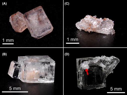
Analyzing carotenoids of snow algae by Raman microspectroscopy and high-performance liquid chromatography
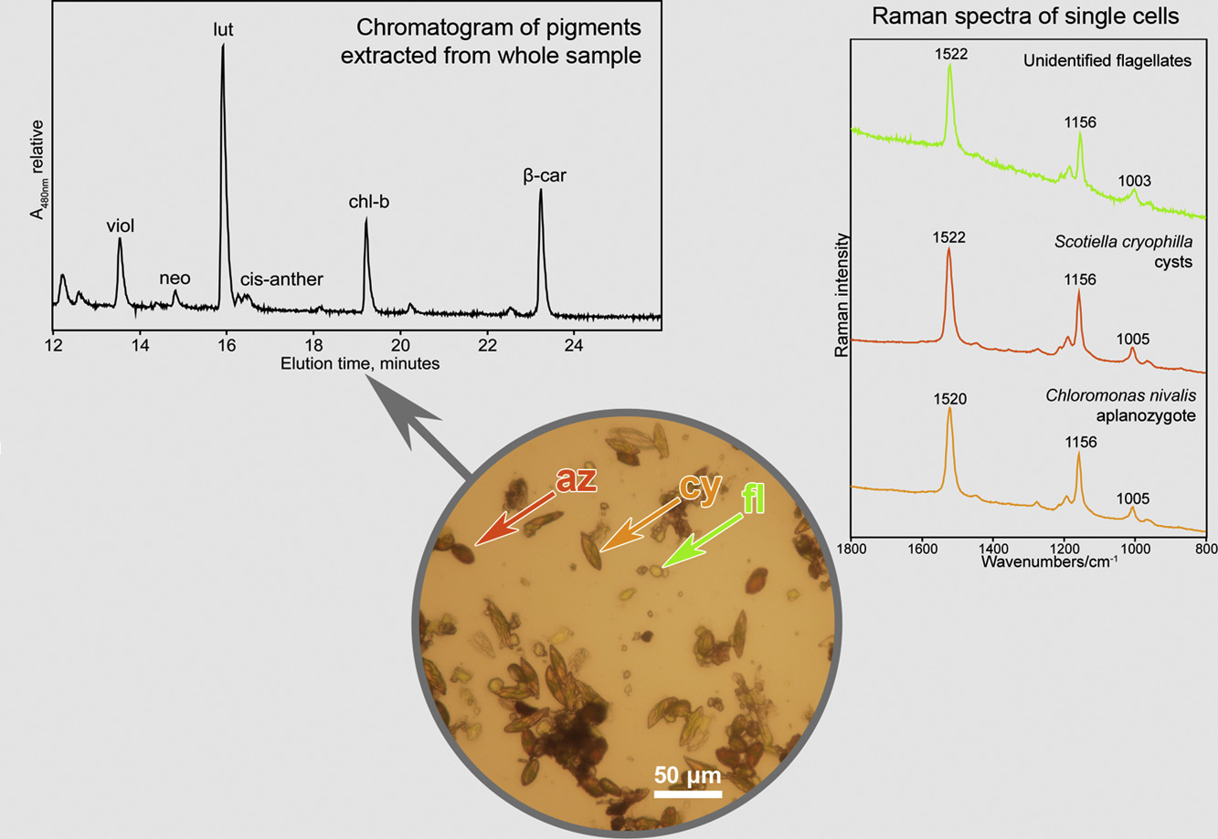
We tested the potential of Raman microspectroscopy to determine carotenoid pigments — both primary (lutein, beta-carotene) and secondary (astaxanthin) carotenoids — in the different species and life-cycle stages of snow algae from the order Chlamydomonadales (Chlorophyta). We compared the performance of Raman spectrometry to a reference method of biological pigment analysis, high-performance liquid chromatography (HPLC). The three main carotenoid Raman bands of the astaxanthin-rich red cysts were located at 1520, 1156 and 1006 cm−1. The shifts (orange aplanozygotes and green motile cells with flagella) in the position of the ν1(C[dbnd]C) Raman band of the polyenic chain is consistent with the expected changes in the ratios of the various carotenoid pigments. Flagellated green cells commonly contain lutein as a major carotenoid, together with minor amounts of β‑carotene and varying amounts of antheraxanthin, violaxanthin and neoxanthin. Aplanozygotes contain mixtures of both primary and secondary carotenoids. In most cases, the ν1(C=C) band is an overlapping set of bands, which is due to the signal of all carotenoid pigments in the sample, and a deconvolution along with the band position shifts (mainly ν1) could be used to characterize the mixture of carotenoids. However, the ability of Raman spectroscopy to discriminate between structurally slightly differing carotenoid pigments or several carotenoids in an admixture in an unknown biological system remains limited. → Link to article at journal webpage.
3. 5. 2018
Using a portable Raman spectrometer to detect carotenoids of halophilic prokaryotes in synthetic inclusions in NaCl, KCl, and sulfates
Cell suspensions of the haloarchaea Halorubrum sodomense and Halobacterium salinarum and the extremely halophilic bacterium Salinibacter ruber (Bacteroidetes) in saturated solutions of chlorides and sulfates (NaCl, KCl, MgSO4·7H2O, K2SO4, and (NH4)Al(SO4)2·12H2O) were left to evaporate to produce micrometric inclusions in laboratory-grown crystals. Raman spectra of these pinkish inclusions were obtained using a handheld Raman spectrometer with green excitation (532 nm). This portable instrument does not include any microscopic tool. Acceptable Raman spectra of carotenoids were obtained in the range of 200–4000 cm−1. This detection achievement was related to the mode of illumination and collection of scattered light as well as due to resonance Raman enhancement of carotenoid signals under green excitation. The position of diagnostic Raman carotenoid bands corresponds well to those specific carotenoids produced by a given halophile. To our best knowledge, this is the first study of carotenoids included in the laboratory in crystalline chlorides and sulfates, using a miniature portable Raman spectrometer. → Link to article at journal webpage.
23. 3. 2018
Raman spectroscopic study of six synthetic anhydrous sulfates relevant to the mineralogy of fumaroles
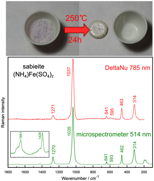
Six anhydrous sulfate phases (millosevichite, mikasaite, efremovite, godovikovite, sabieite, and steklite) were prepared by heating in the laboratory and analyzed using a laboratory Raman microspectrometer and a miniaturized Raman spectrometer. Raman spectra are reported, and assignments of fundamental vibrational modes are proposed. Experience with the miniature Raman instrument is discussed → Link to article at journal webpage.
18. 10. 2017
Burning coal spoil heaps as a new habitat for the extremophilic red alga Galdieria sulphuraria
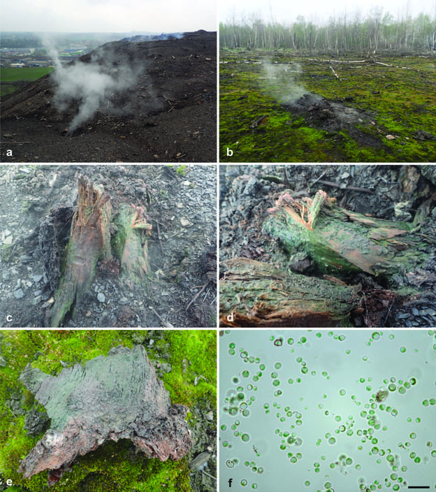
Galdieria sulphuraria (Cyanidiales) is a worldwide acclaimed thermoacidophilic red microalga with a limited distribution due to special conditions required for growth and metabolism. Until now, the alga was almost exclusively restricted to acid geothermal environments around the world. However, we have found this species on the surface of a burning coal spoil heap in central Europe. It is the first record of G. sulphuraria in this type of habitat. A rbcL phylogeny confirmed that the population of this extremophile belongs to the continental European lineage and we consider Italian geothermal sites as a potential source of Czech G. sulphuraria. The dispersal of unicellular red microalgae is far from fully understood and the discovery of Galdieria in another region of Europe on a relatively newly established anthropogenic site allows us to understand better the distribution patterns and dispersal abilities of this ecologically important algal group. In addition, we have also analyzed the phylogenetic position of Galdieria strain CCALA 965 isolated from a highly acidic site without geothermal activity in the Czech Republic and confirmed it to belong to the species G. phlegrea, until now known only from Italy. → Link to article at journal webpage.