Exobiology Group
Institute of Geochemistry, Mineralogy and Mineral Resources
Faculty of Science, Charles University
We are interested in the applications of Raman spectroscopy in the fields of exobiology, geochemistry or geoscience in general. The topics of our research include:
Biomarkers
Detecting and discriminating biomarkers as well as understanding their behaviour in the geological record is a an important task for geoscience and geomicrobiology. There is a need of a new step in the following directions a) emerging types of colonisations by extremophiles, b) new biomarkers of high relevance for Mars exobiology c) new analytical developments. Raman spectroscopy being involved in the exobiological field studies on Earth, in a perspective of including a miniature Raman instrument in the flyby of the next mission to Mars (as in the frame of ExoMars).
Carbons
The transformation of organic matter during diagenesis and metamorphism can lead under certain conditions to the formation of solid residues. These residues can occur disseminated or accumulated in very diverse settings and mineral associations. Obtaining knowledge about the structure and microtexture of these residues is helpful to solve some of the major questions of geoscience i.e. how about the biogenicity of the geological residue, how about the type of precursor biomass or what were the conditions and timing of the geological transformation processes.
Extremophiles
Extremophiles originating from various habitats (hypersaline pools, polar lakes, snow, hot springs) as well as strains obtained from reference collections can be cultivated to produce sufficient biomass for complex analytical investigations focused onto pigments, osmotic solutes and lipids. We conduct direct field investigations of accumulated microbial biomass/mats/endoliths. Direct field analyses are carried-out using mobile/miniaturized instrumentation allows us to collect basic detection information to be gathered fast and directly on outcrops.
Minerals
Application of miniaturized/handheld Raman instrumentation for the studies at geological sites (analogues) that allow us to assess and address the challenges of in situ spectroscopic research with focus especially on more or less hydrated minerals of importance for the Martian exobiology, such as sulfates.
Abstract of previously published works
13. 1. 2019
Analyzing carotenoids of snow algae by Raman microspectroscopy and high-performance liquid chromatography
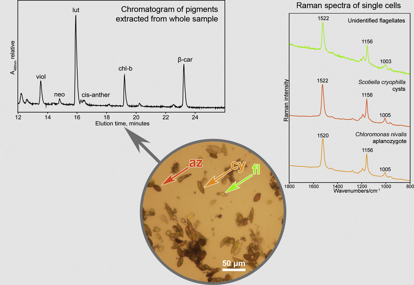
We tested the potential of Raman microspectroscopy to determine carotenoid pigments — both primary (lutein, beta-carotene) and secondary (astaxanthin) carotenoids — in the different species and life-cycle stages of snow algae from the order Chlamydomonadales (Chlorophyta). We compared the performance of Raman spectrometry to a reference method of biological pigment analysis, high-performance liquid chromatography (HPLC). The three main carotenoid Raman bands of the astaxanthin-rich red cysts were located at 1520, 1156 and 1006 cm−1. The shifts (orange aplanozygotes and green motile cells with flagella) in the position of the ν1(C[dbnd]C) Raman band of the polyenic chain is consistent with the expected changes in the ratios of the various carotenoid pigments. Flagellated green cells commonly contain lutein as a major carotenoid, together with minor amounts of β‑carotene and varying amounts of antheraxanthin, violaxanthin and neoxanthin. Aplanozygotes contain mixtures of both primary and secondary carotenoids. In most cases, the ν1(C[dbnd]C) band is an overlapping set of bands, which is due to the signal of all carotenoid pigments in the sample, and a deconvolution along with the band position shifts (mainly ν1) could be used to characterize the mixture of carotenoids. However, the ability of Raman spectroscopy to discriminate between structurally slightly differing carotenoid pigments or several carotenoids in an admixture in an unknown biological system remains limited. → Link to article at journal webpage.
3. 5. 2018
Using a portable Raman spectrometer to detect carotenoids of halophilic prokaryotes in synthetic inclusions in NaCl, KCl, and sulfates
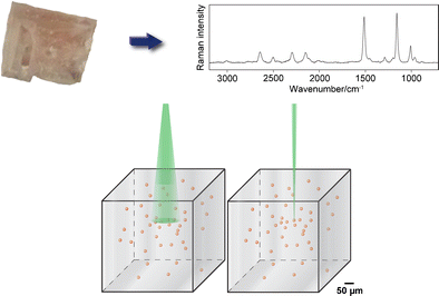
Cell suspensions of the haloarchaea Halorubrum sodomense and Halobacterium salinarum and the extremely halophilic bacterium Salinibacter ruber (Bacteroidetes) in saturated solutions of chlorides and sulfates (NaCl, KCl, MgSO4·7H2O, K2SO4, and (NH4)Al(SO4)2·12H2O) were left to evaporate to produce micrometric inclusions in laboratory-grown crystals. Raman spectra of these pinkish inclusions were obtained using a handheld Raman spectrometer with green excitation (532 nm). This portable instrument does not include any microscopic tool. Acceptable Raman spectra of carotenoids were obtained in the range of 200–4000 cm−1. This detection achievement was related to the mode of illumination and collection of scattered light as well as due to resonance Raman enhancement of carotenoid signals under green excitation. The position of diagnostic Raman carotenoid bands corresponds well to those specific carotenoids produced by a given halophile. To our best knowledge, this is the first study of carotenoids included in the laboratory in crystalline chlorides and sulfates, using a miniature portable Raman spectrometer. → Link to article at journal webpage.
23. 3. 2018
Raman spectroscopic study of six synthetic anhydrous sulfates relevant to the mineralogy of fumaroles
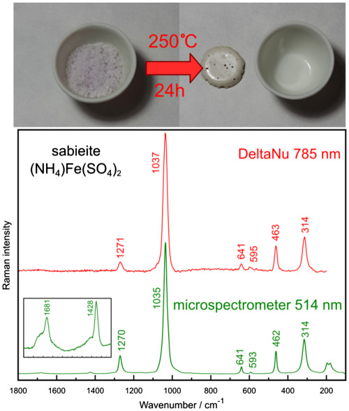
Six anhydrous sulfate phases (millosevichite, mikasaite, efremovite, godovikovite, sabieite, and steklite) were prepared by heating in the laboratory and analyzed using a laboratory Raman microspectrometer and a miniaturized Raman spectrometer. Raman spectra are reported, and assignments of fundamental vibrational modes are proposed. Experience with the miniature Raman instrument is discussed → Link to article at journal webpage.
18. 10. 2017
Burning coal spoil heaps as a new habitat for the extremophilic red alga Galdieria sulphuraria
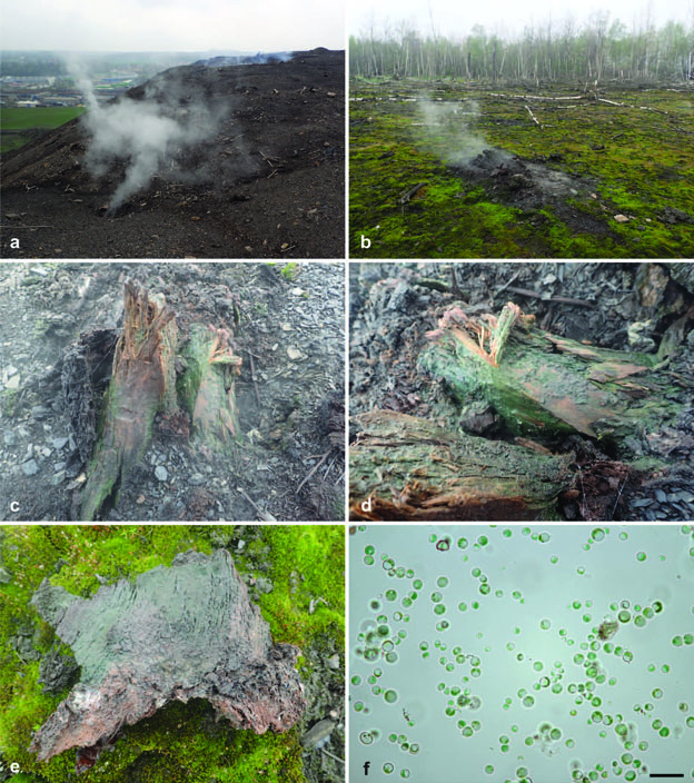
Galdieria sulphuraria (Cyanidiales) is a worldwide acclaimed thermoacidophilic red microalga with a limited distribution due to special conditions required for growth and metabolism. Until now, the alga was almost exclusively restricted to acid geothermal environments around the world. However, we have found this species on the surface of a burning coal spoil heap in central Europe. It is the first record of G. sulphuraria in this type of habitat. A rbcL phylogeny confirmed that the population of this extremophile belongs to the continental European lineage and we consider Italian geothermal sites as a potential source of Czech G. sulphuraria. The dispersal of unicellular red microalgae is far from fully understood and the discovery of Galdieria in another region of Europe on a relatively newly established anthropogenic site allows us to understand better the distribution patterns and dispersal abilities of this ecologically important algal group. In addition, we have also analyzed the phylogenetic position of Galdieria strain CCALA 965 isolated from a highly acidic site without geothermal activity in the Czech Republic and confirmed it to belong to the species G. phlegrea, until now known only from Italy. → Link to article at journal webpage.
8. 6. 2017
Applying portable Raman spectrometers for field discrimination of sulfates: Training for successful extraterrestrial detection
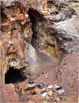
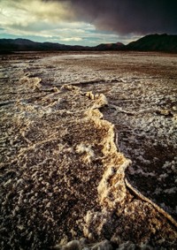
The presence of sulfates containing Mg, Fe, and Ca (in several hydration states) has been confirmed on Mars from data acquired by both orbital and in-situ measurements. It has been suggested that the sulfates on Mars originated deep the past from the evaporation of water at accumulation points on the Martian surface. Consequently, they can be seen as highly relevant to any search for the presence of extraterrestrial life. Miniaturized Raman spectrometers (which permit analysis of the mineralogical and molecular composition of a specimen) will be included in rovers on future planetary exploration missions. Portable miniature Raman spectrometers operating in the nearinfrared (785 nm) and in the green (532 nm, currently selected for the ExoMars mission) were used on-site to estimate the mineralogical compositions of sulfate weathering crusts at Valachov (Bohemian Massif, Czech Republic). Spectra of sufficient quality were quickly obtained, and the characteristic shifts of the sulfate ν1 band of the minerals allowed for the discrimination of several sulfates. Superficial crusts contained gypsum, jarosite, rozenite, fibroferrite, magnesiocopiapite, and melanterite as major components. The current testing has confirmed the favorable performance of handheld instruments for the discrimination of structurally similar sulfates of relevance for studies on Mars. → Link to article at journal webpage.
11. 4. 2017
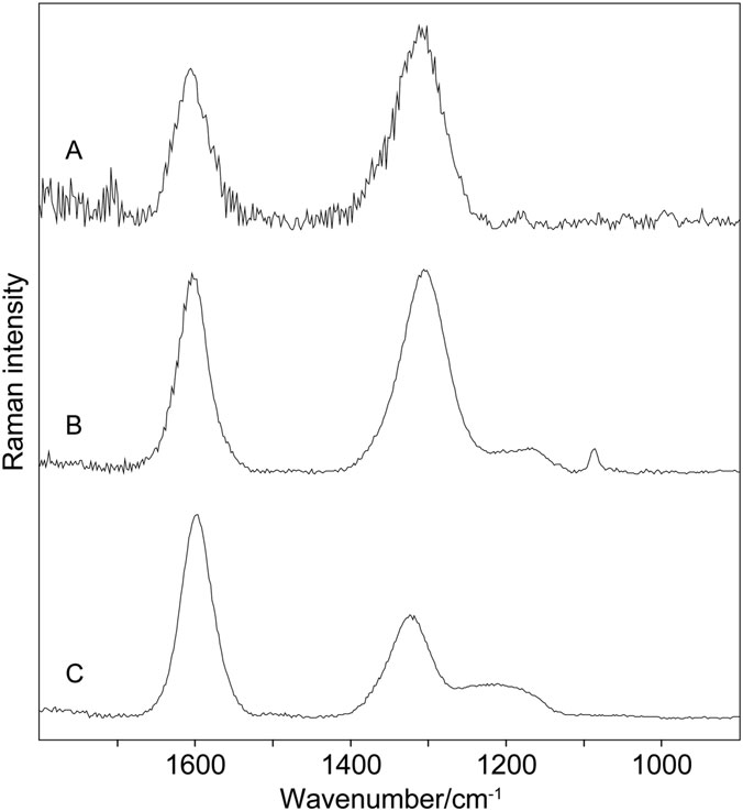
Raman microspectrometric study of pigments in melanized fungi from the hyperarid Atacama desert gypsum crust
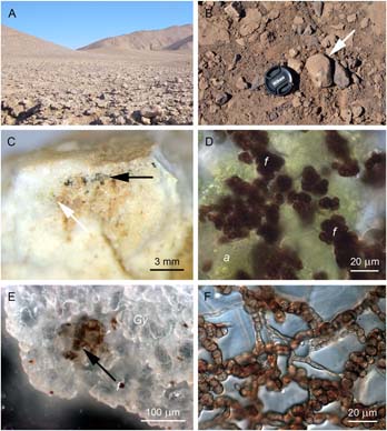
The Atacama desert is amongst the oldest and driest deserts in the world, and its hyperarid core is described as ‘the most barren region imaginable’. In this study, we describe for the first time the endolithic microbial communities composed by eukaryotic microorganisms, such as melanized fungi and algae, colonizing the interior of gypsum crusts collected in the hyperarid zone of the Atacama desert. Melanin pigments present in fungi cell walls have been studied by Raman microspectroscopy. Analyses have been performed directly in the gypsum crust sustaining the colonisation of fungi and algae, on the thin sections and on the cultivated culture of the fungi from Neocatenulostroma genus. Raman spectroscopic signature of melanin has been clearly observed in all the sample types. Two broad bands located at wavenumbers 1605–1580 cm-1 and around 1350 cm-1 have been detected in all the studied samples, with slight variations in the appearance of other bands or shoulders of smaller intensity documenting either the changes in melanin composition or other biomolecules bound to melanin. Blue and green lasers (445, 514 and 532 nm) have been shown as favourable excitation for the study of melanins as the longer wavelengths induced adverse effects that mask the Raman signal. Effect of laser power level on the resulting spectra has also been discussed. Algal carotenoid pigment has been detected in isolated regions of the sample by the Raman bands of 1520, 1155 and 1002 cm-1. The strong Raman signal of fungal melanin that has been detected in the host rock from the locality characterized by extreme conditions such as Atacama desert could be considered in the discussion about feasible biomarker molecules; however, low specificity of the broad Raman signature and similarity to the spectrum of amorphous carbon must be also taken into account. → Link to article at journal webpage.
10. 2. 2017
Obtaining Raman spectra of minerals and carbonaceous matter using a portable sequentially shifted excitation Raman spectrometer – a few examples
We present examples of application of a recently developed portable sequentially shifted excitation Raman spectrometer for identifying three series minerals and carbonaceous matter. Those include compounds of relevance for the fields of geobiology and exobiology: sulfates and carbonates, organic minerals, and carbonaceous matter. It is demonstrated that unambiguous obtaining of Raman spectra can be achieved fast and with the gain in eliminating potential fluorescence features. → Link to article at journal webpage.
8. 12. 2016
Colonization of Snow by Microorganisms as Revealed Using Miniature Raman Spectrometers—Possibilities for Detecting Carotenoids of Psychrophiles on Mars?

We tested the potential of a miniaturized Raman spectrometer for use in field detection of snow algae pigments. A miniature Raman spectrometer, equipped with an excitation laser at 532 nm, allowed for the detection of carotenoids in cells of Chloromonas nivalis and Chlamydomonas nivalis at different stages of their life cycle. Astaxanthin, the major photoprotective pigment, was detected in algal blooms originating in snows at two alpine European sites that differed in altitude (Krkonoše Mts., Czech Republic, 1502m a.s.l., and Ötztal Alps, Austria, 2790m a.s.l.). Comparison is made with a common microalga exclusively producing astaxanthin (Haematococcus pluvialis). The handheld Raman spectrometer is a useful tool for fast and direct field estimations of the presence of carotenoids (mainly astaxanthin) within blooms of snow algae. Application of miniature Raman instruments as well as flight prototypes in areas where microbes are surviving under extreme conditions is an important stage in preparation for successful deployment of this kind of instrumentation in the framework of forthcoming astrobiological missions to Mars. → Link to article at journal webpage.
31. 10. 2016
A unique application of handheld Raman instruments for a quick identification of large quantities of gemstones on a Ring Monstrance from Prague's Loreto's treasury

A miniature lightweight portable Raman spectrometer and a palm-sized device allow for fast and unambiguous detection of common gemstones mounted in complex jewels. Here, complex religious artefacts and the Ring Monstrance from the Loreto treasury (Prague, Czech Republic; eighteenth century) were investigated. These discriminations are based on the very good correspondence of the wavenumbers of the strongest Raman bands of the minerals. Very short laser illumination times and efficient collection of scattered light were sufficient to obtain strong diagnostic Raman signals. The following minerals were documented: quartz and its varieties, beryl varieties (emerald), corundum varieties (sapphire), garnets (almandine, grossular), diamond as well as aragonite in pearls. Miniature Raman spectrometers can be recommended for common gemmological work as well as for mineralogical investigations of jewels and cultural heritage objects whenever the antiquities cannot be transported to a laboratory. → Link to article at journal webpage.
26. 7. 2016
A vibrational spectroscopic study of an interesting red pigment prodigiosin has been published.
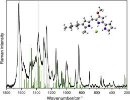
Prodigiosin is a pink colored tripyrrole pigment which occurs naturally as a biosynthetic product of Serratia marcescens. The direct acquisition of Raman spectra of this compound from fresh cultures of S. marcescens was found to be not possible even with a range of different excitation conditions and common instrumental settings. Therefore, IR and Raman spectroscopic data are given here for pure prodigiosin hydrochloride. DFT calculations of possible isomers were undertaken to better understand the spectral differences potentially arising from the different structures. Comparison of experimental and theoretical data shows a good correspondence with the selected molecular structure. The spectroscopic data obtained from prodigiosin identify spectral biomarker signatures which can complement information and databases obtained for other pigments considered as biosignatures in the fields of geobiology and exobiology. → Link to article at journal webpage.
12. 12. 2014
Special issue of Philosophical Transactions on Raman spectroscopy applications in Mars exobiology
The special issue published in Royal Transactions A and edited by Jan Jehlička and Howell G. M. Edwards covers the applications of Raman spectroscopy to subjects of relevance for Mars exobiology. The papers focus on biomarkers and their Raman spectroscopic detection in the frame of extremophile-rich ecosystems and relevant rock representatives, including both laboratory and theoretical studies. Reports on successful field applications of miniaturized Raman spectrometers and Mars prototypes for biomarkers and mineral identification will complement laboratory research. This topic is currently receiving a great deal of attention due to the forthcoming ESA/Roscomos 2018 ExoMars mission and the proposed NASA 2020 mission, both of which will use Raman spectroscopic instruments in the search of life. PDF